Shoulder Tendinopathy Treatment
Introduction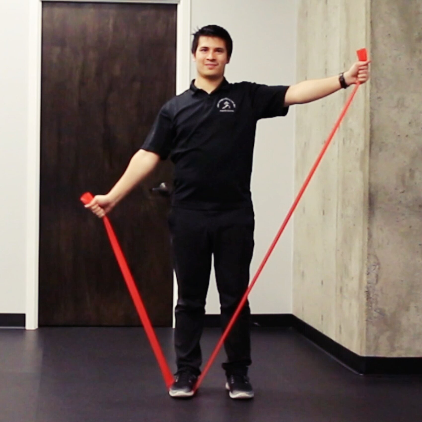
Tendinopathy is a common cause of pain in the shoulder. Although it is a common condition, there is a lot of confusion about tendon pain. From whether inflammation causes tendon pain to which exercises are most effective. This post will cover:
- the mechanism of how shoulder tendinopathy develop,
- how to diagnose shoulder tendinopathy,
- whether imaging is indicated,
- and which treatments are most effective for shoulder tendinopathy.
Pathology of Tendinopathy
The pathophysiology of tendon pain has transitioned over the previous few decades. The current model of tendon pain is the continuum model purposed by Jill Cook and Craig Purdam. In the continuum model, there are three stages of tendinopathy:
- reactive tendinopathy,
- tendon dysrepair,
- and degenerative tendinopathy.
In clinical practice, these are divided into two stages:
- reactive-early tendon dysrepair tendinopathy
- and late tendon dysrepair-degenerative tendinopathy.
A reactive-early tendon dysrepair tendinopathy occurs after a sudden increase in physical activity. The sudden spike in activity leads to activation of tenocytes. This activation of tenocytes then causes swelling and mild separation of the tendon fibers. These changes in the early phase of tendinopathy are generally reversible with adequate rest.Prolonged overloading of the tendon can lead to a late tendon dysrepair-degenerative tendinopathy. These individuals usually have a history of repeated bouts of tendon pain. The pain during each bout increases as the amount of load on the tendon increases. In this stage of tendinopathy, there is extensive matrix and collagen disorganization. Neovascularization of the tendon also occurs in degenerative tendons. The formation of blood vessels in the tendon has been implicated as a cause of persistent pain.The prolonged overload on the tendon can also cause cell apoptosis. This cell apoptosis may be seen as either a full or partial thickness tear of the rotator cuff. The changes in a degenerative tendinopathy are generally not reversible. But this does not mean that someone will have persistent pain or will never be able to return to activity. The reason is because structure of the tendon is poorly correlated to pain.
Physical Examination of the Shoulder
The purpose of a physical examination is to stress tissues to determine the cause of pain. For these physical exams to be useful in clinical practice, they need to be both specific and sensitive for the condition. This means they are positive when the condition is present (specific) and negative when not present (sensitive). The benchmark value for orthopedic testing is 80% for both specificity and sensitivity.A systematic review of orthopedic testing for the shoulder concluded "the use of any single shoulder physical exmaination test to make a diagnosis cannot be unequivocally endorsed." But the combination of:
- age greater than 39,
- a positive painful arc,
- and self reported popping or clicking,
was the most useful to diagnose a supraspinatus tendinopathy. The presence of 2 out of the 3 factors resulted in 75% sensitivity and 99% specificity. While the presence of all 3 factors had a 38% sensitivity and 99% specificity. However, this combination fails to reach the 80% benchmark.In another study for bicipital tendinopathy, the combination of:
- Yergason's test,
- Speed's test,
- and popeye's sign,
had a sensitivity of 73% and specificity of 58%. But again, falls short of the recommended specificity and sensitivity .Even though physical examination cannot isolate specific tissues, they can still be valuable. The pain provoking movements found in testing can be combined with the shoulder symptom modification procedure (SSMP) to guide treatment. The SSMP is an approach developed by Jeremy Lewis to assess the shoulder and guide rehab. It begins with changing the position of the thoracic spine and reassessing a pain provoking movement. The procedure then proceeds to different body regions to assess their influence on the shoulder pain. To read more on the shoulder symptom modification procedure, here is a link to the article.
Imaging for Shoulder Tendinopathy
Structural abnormalities have been commonly attributed as the source of pain in the shoulder. Rotator cuff tears, bone spurs, and osteoarthritis are common diagnoses for shoulder pain. The association of these structural pathologies to pain frequently leads to imaging to assess the shoulder.A study in BJSM found that MRI, MRA, and ultrasound (US) were all effective in detecting rotator cuff tears. The systematic review found MRI, MRA, and US had 90% sensitivity and specificity for full thickness rotator cuff tears. The study also found the sensitivity and specificity was similar for partial thickness tears.There is no doubt that these imaging techniques are helpful in detecting structural pathology. But the clinical significance of these structural pathologies has recently been questioned. This is due to the high rate of structural pathology found in those without shoulder pain.
- A study of 42 workers with shoulder impingement and 31 age-matched workers with no shoulder impingement. An MRI was then performed of the shoulders in both groups . The study found that 55% of the subjects in the impingement group had a pathologic supraspinatus tendon. While in the control group, 52% of the subjects also had a pathologic supraspinatus tendon.
- Another study performed an ultrasound on 90 subjects with no history of shoulder pain. The study found over 50% of the subjects over 70 years old had a partial or full thickness tear. This increased to over 80% in subjects over 80 years old. Of note, this study also did not find a statistical difference between rotator cuff pathology and hand dominance.
- In another study looked at 51 male subjects without a history of shoulder pain. This study found shoulder abnormalities in 96% of the asymptomatic individuals. The most common shoulder abnormality was subacromial/subdeltoid bursa thickening (78%). Other shoulder abnormalities found were:
- acromioclavicular joint osteoarthritis (65%),
- supraspinatus tendinosis (39%),
- rotator cuff partial thickness tear (22%),
- and supraspinatus full thickness tear (9.8%).
These structural pathologies may be the cause of pain in the shoulder but should be considered with caution. The above studies concluded that these structural changes are likely part of the normal aging process, not necessarily pain.The issue with linking these structural changes to pain is that they frequently need surgery to correct. Since these changes are common in pain free subjects, this may lead to unnecessary surgery. So it is crucial whenever imaging is performed on the shoulder to correlate the patient's symptoms with the image, and not the other way around.
Reactive Tendinopathy/Early Tendon Dysrepair Treatment
The emphasis in the treatment of reactive tendinopathy should be on load management. Activities that place a fast, high load on the tendon (ie. plyometrics) or activities that cause compression of the tendon should be temporarily avoided. These activities may include push ups, bench press, dips, and overhead pressing. Many activities in sports may also need to avoided because of their ballistic nature. Common examples are throwing and swinging motions.Reducing the total amount of loading placed on the tendon is necessary for a reactive tendinopathy. Yet, completely off-loading the tendon is not advised because it may lead to stress shielding. This will lead to a weaker tendon and an increased risk of re-injury. Instead a period of relative rest should be the focus of load management.A period of relative rest is achieved by altering the intensity, duration, and frequency of loading on the tendon. To determine how much load a tendon can tolerate, the general rule is:
- pain less than 3/10 during activity,
- no increased pain 24 hours after exercise,
- and pain doesn’t interfere with sleep.
Isometric exercises have emerged as the preferred treatment method for pain management. For isometric exercises to produce an analgesic effect, a heavier resistance is necessary. The recommendation for the shoulder is to use a weight 50% of maximal strength for 3 reps of 30 seconds.Additionally, manual therapy can be an adjunct therapy to exercise. A systematic review in JOSPT found manual therapy resulted in a significant reduction in pain but the effect on function was not clear. As a result, manual therapy should not be used as an isolated treatment. Instead, it can be used as an adjunct therapy to support exercise therapy.
Late Tendon Dysrepair/Degenerative Tendinopathy Treatment
The focus in degenerative tendinopathy is to increase tendon capacity through loading. Load management is also still an important consideration. But to improve tendon capacity, isotonic exercises are essential to build more tendon.Unfortunately, there does not appear to be an optimal loading strategy for tendinopathy. The standard 3 sets of 10 reps is the most common recommendation in the literature. Another loading option is 5 sets of 5-6 reps which may allow for the use of higher weights with less fatigue. For now, the decision on which loading strategy to use will depend on the individual patient.Pain experienced during exercise is a common limitation to loading. The fear is pain means the loading is causing damage to the tendon. Yet in many of the loading programs, pain is acceptable during rehabilitation. The reason is the tendon needs load to stimulate the tenocytes to build more tendon.A systematic review in BJSM found that pain during exercise may have small short term benefits over pain-free exercises. The study also found that pain was not associated with negative treatment outcomes. While pain may not be a desired experience during treatment, short term discomfort may lead to longer term benefits.Patients with degenerative tendinopathy can become frustrated by the persistent pain. This leads many to inquire whether medications and injections are appropriate for care. Corticosteroids and NSAIDs can provide short term pain relief but can cause longer term consequences. Both corticosteroids and NSAIDs are associated with delayed soft tissue healing. Corticosteroid injections are also associated with increased reoccurrence rates. These outcomes are the opposite of the desired treatment goals. The goal of therapy is to increase tissue capacity and reduce re-injury. Corticosteroids and NSAIDs are thus not advised in the treatment of degenerative tendinopathy.
Conclusion
Shoulder tendinopathy is an overload condition, not driven by inflammation. An acute spike in activity can cause a reactive tendinopathy. The primary treatment of reactive tendinopathy is load management. With continued overload, a degenerative tendinopathy can develop. A progressive exercise program is the primary strategy for a degenerative tendinopathy.Manual therapy may be used as an adjunct therapy for tendinopathy. Other treatments such as injections and NSAIDs may play a small therapeutic role. But none of these treatments should be used in place of a progressive exercise program.
References:
- Cook, J L, and C R Purdam. “Is Tendon Pathology a Continuum? A Pathology Model to Explain the Clinical Presentation of Load-Induced Tendinopathy.” British Journal of Sports Medicine, vol. 43, no. 6, 2008, pp. 409–416., doi:10.1136/bjsm.2008.051193.
- Coombes, Brooke K., et al. “Effect of Corticosteroid Injection, Physiotherapy, or Both on Clinical Outcomes in Patients With Unilateral Lateral Epicondylalgia.” Jama, vol. 309, no. 5, June 2013, p. 461., doi:10.1001/jama.2013.129.
- Desjardins-Charbonneau, Ariel, et al. “The Efficacy of Manual Therapy for Rotator Cuff Tendinopathy: A Systematic Review and Meta-Analysis.” Journal of Orthopaedic & Sports Physical Therapy, vol. 45, no. 5, 2015, pp. 330–350., doi:10.2519/jospt.2015.5455.
- Frost, Poul, et al. “Is Supraspinatus Pathology as Defined by Magnetic Resonance Imaging Associated with Clinical Sign of Shoulder Impingement?” Journal of Shoulder and Elbow Surgery, vol. 8, no. 6, 1999, pp. 565–568., doi:10.1016/s1058-2746(99)90090-3.
- Hegedus, Eric J, et al. “Which Physical Examination Tests Provide Clinicians with the Most Value When Examining the Shoulder? Update of a Systematic Review with Meta-Analysis of Individual Tests.” British Journal of Sports Medicine, vol. 46, no. 14, July 2012, pp. 964–978., doi:10.1136/bjsports-2012-091066.
- Lewis, J S. “Rotator Cuff Tendinopathy/Subacromial Impingement Syndrome: Is It Time for a New Method of Assessment?” British Journal of Sports Medicine, vol. 43, no. 4, Jan. 2009, pp. 259–264., doi:10.1136/bjsm.2008.052183.
- Lewis, Jeremy, et al. “Rotator Cuff Tendinopathy: Navigating the Diagnosis-Management Conundrum.” Journal of Orthopaedic & Sports Physical Therapy, vol. 45, no. 11, 2015, pp. 923–937., doi:10.2519/jospt.2015.5941.
- Littlewood, Chris, et al. “Exercise for Rotator Cuff Tendinopathy: a Systematic Review.” Physiotherapy, vol. 98, no. 2, 2012, pp. 101–109., doi:10.1016/j.physio.2011.08.002.
- Naredo, E. “Painful Shoulder: Comparison of Physical Examination and Ultrasonographic Findings.” Annals of the Rheumatic Diseases, vol. 61, no. 2, Jan. 2002, pp. 132–136., doi:10.1136/ard.61.2.132.
- Frost P, Andersen JH, Lundorf E. Is supraspinatus pathology as defined by magnetic resonance imaging associated with clinical sign of shoulder impingement? J Shoulder Elbow Surg 1999;8:565-568.
- Milgrom C, Schaffler M, Gilbert S, et al. Rotator-cuff changes in asymptomatic adults. The effect of age, hand dominance and gender. J Bone Joint Surg Br 1995;77:296-298
- Rees, J D, et al. “Tendons: Time To Revisit Inflammation?” British Journal of Sports Medicine, vol. 47, no. 9, Oct. 2013, doi:10.1136/bjsports-2013-092459.9.
- Rio, Ebonie, et al. “The Pain of Tendinopathy: Physiological or Pathophysiological?” Sports Medicine, vol. 44, no. 1, Dec. 2013, pp. 9–23., doi:10.1007/s40279-013-0096-z.
- Roy, Jean-Sébastien, et al. “Diagnostic Accuracy of Ultrasonography, MRI and MR Arthrography in the Characterisation of Rotator Cuff Disorders: a Systematic Review and Meta-Analysis.” British Journal of Sports Medicine, vol. 49, no. 20, Nov. 2015, pp. 1316–1328., doi:10.1136/bjsports-2014-094148.
- Smith, Benjamin E, et al. “Should Exercises Be Painful in the Management of Chronic Musculoskeletal Pain? A Systematic Review and Meta-Analysis.” British Journal of Sports Medicine, vol. 51, no. 23, Aug. 2017, pp. 1679–1687., doi:10.1136/bjsports-2016-097383.